Tagged: anatomy and physiology of wine tasting
Sight, Anatomy and Physiology of Wine Tasting – Part 2 Sight Receptors
Wine is sunlight, held together by water. – Galileo
sense one in wine tasting – SIGHT, as appeared on https://wine4soul.com/2012/10/28/vision-anatomy-and-physiology-of-wine-tasting/ continues…
Physiologically, sight is initiated when reflected light of different wave lengths: colour, hit the eye.

We have the bottle before us, the glass is filled (no more than a third up), the reflected light in different colours hits the external surface of the eyeball: The Cornea, travels through the lens and inwards through the Vitreous Humor, to hit the Retina, the innermost layer of the eye ball. The Retina consists of nerve tissue, Photo-receptors that sense the light entering the eye, and start translating it as the images of our vision in the form of shape and colour in the brain.
The Physiology of sight

Photoreceptors are specialty cells in the Retina, they allow us to see shapes, colors and the combination of both, something we all take for granted.
How is it done? The retina contains 2 kinds of photoreceptors:
 1. Rod cells: These function only in dim light and are blind to colour. Only the highest-intensity output gets through; so contrast and visual definition are improved.
These receptors enter into function when we enter a dark cellar full of wine wonders, or when we stroll down the vineyard at night (with or without our lover, better with!)
2. Cone cells: The other kind of photoreceptor cell. There are three different types of cones, sensitive to different light wave lengths (Red Green & Blue). The cones operate in bright light and are responsible for high acuity vision, as well as ability to “see” colour.
Rods and cones form an uneven mosaic within the retina; there are 10 times more rod cells than cones. Rods are concentrated at the outer edges of the retina. There are approximately 130 million rod cells in the human retina. Rod cells are almost entirely responsible for peripheral and night vision. They are 100 times more sensitive to a single photon than cones so rods require less light to function than cones and allow us to see in the dark. Single Rod cells collect and amplify light signals. However, this convergence comes at a cost to visual acuity / resolution, because the accumulated information
  from several cells simultaneously is less accurate than information from each rod cell individually. But that will have to do since we look at wine under good light conditions and not in the dark.
In the retina’s center – fovea, cones are highly concentrated 5-10 times more than on the rest of the retinal surface, this area is described by Nobel Prize winner Jeremy Nathans as: “the most valuable square millimeter of tissue in the body.”

The first kind of cone responds to red colour (light of Long wavelengths – L around 564–580 nm); The second type responds to green colour (Medium wavelength – M, 534–545 nm), The third type responds to blue colour (Short wavelength – S, 420–440 nm), The difference in the signals received from the three cone types in varying degree of stimulus strength which allows the brain to perceive all possible colours. The brain combines the information from each type of receptor to give rise to different perceptions of different wavelengths of light and ultimately the correct colour.
 Wine comes in a wide variety of colours shades and hues. It is these receptors and the wonders of the final interpretation of these signals into what we call sight, in our brain, will allow us to distinguish between different grape varieties, wines from different regions, wines in different state of evolution and wine making methods just by mere sight, with no other senses involvement.
Several theories explain the mechanism of colour vision, Helmholtz’s trichromatic theory & Hering’s opponent process theory, they differ on the exact point colour processing actually begins, either within the receptor cells in the retina or slightly behind it, at the level of retinal ganglion cells and beyond. Visual information is then sent to the brain from retinal ganglion cells via the optic nerve to the optic chiasm: a point where the two optic nerves meet and cross each other. Information from one visual field crosses to the other side of the brain to the visual cortex.
Cone cells allow us to stabilize the colour constancy of an object, so when we look at red wine we preserve an ability to see the true colours of our object for instance red wine in different hues and shades.
clarity:
The wine clarity is wine easily examined at a slight tilt under clear light conditions either with the background of a white paper or a well lit background. And brightness is reflected from it. Fresh wine should have a clear spark ‘sneaky’ kind of wink and it looks sleek and shiny.
Clarity is graded to 3-4 levels:
Brilliant or Crystalline: perfect transparency; the surface of the wine reflects the light with a sparkle.
Clear: normal state of clarity
Dull: a partial lack of luster
Cloudy or slushy: with or without suspended particles visible to the naked eye

Wine, whatever its category should be clear, perfectly transparent and free of foreign deposits or suspended particles, most suspended particles are wine deposits and are not associated with wine faults. Signs of cloudiness may indicate a defect. A fine wine of any color at its prime should be not only clear but also bright with a luminous quality.
Type of Wine – Obviously, different types of wines will have dramatically different wine colour..White wines tend toward the more clear yellow and gold end of the spectrum while red wines can vary from light red to deep purple. Rosé wines are somewhere in between. Additionally, your expectation of what a wine should look like depends on the type of wine in question. For example, while many Cabernet Sauvignon, 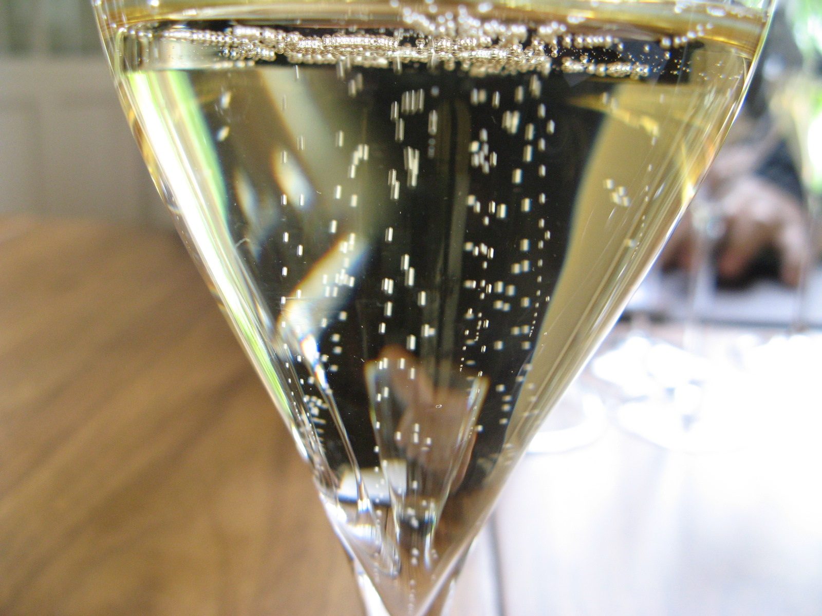 based wines will be dark purple or even close to opaque, Pinot Noir-based wines tend to be lighter with less depth of color and a lighter hue.
Is the color of the wine appropriate for the type of wine you taste? You will learn this as you go along and get more experience with different types of wines.
The COLOURS of WINE
Colour is simply light of different wavelengths and frequencies that we can actually see and is made up from photons, reflection of light from the wine (as our object) is what we see in form, shape, and colour with its inner diversity of different hues and shades. .
Wine colours, originate from the grape’s skin. Grape juice from red or white varieties is usually transparent (clear to cloudy). Anthocyanins are the chemical compounds that give wine white or red its colour, or pigment.
Different “exposure” to grape treatments like: amount and type of crushing, which exerts colors into the liquid. Changes in temperature, contact of broken skin with the juice, exposure to oxygen, fermentation with or without the skins (lees), length of fermentation, type of tanks: barrel or steel, etc. All of these factors change and affect the wine’s colour, which keeps changing even after bottling, as aging affects the depth and hue of the basic color of each wine. Different grape varieties contribute different hues of white yellow or red as described below.
WHITE WINES
 Polyphenols contribute to the yellow colour of white wines, phenols concentration in different grape
 variety varies: level of phenols in the Riesling grape is very low hence they appear almost transparent, Chardonnay on the other hand due to high phenolic concentration will appear darker : yellow lemony colour. Apart from phenols, maturity level of the grape will also affect the colour. The riper the grape, the darker shades of yellows in white wines.
The colors of the wine can vary strongly depending on age, concentration and wine making techniques. Colour of white wines deepens with age, tending toward full straw or pale gold. More mature dry wines, particularly if aged in wood, take on rich golden tones, sometimes even with hints of copper or brass. Brown hues are a sign of over oxidation, (a defect in wine), but in certain fortified wines such as Marsala, it is a normal feature. Hints of red in a white wine are usually indications of a fault.
White Wine Colours:
The grapes and wines below, usually exhibit the listed colors.
Clear with a greenish tint-: Indicative of young wines with residual chlorophyll, mainly from cold growing regions: Chardonnay from Chablis, German Rieslings, and young New Zeeland Sauvignon Blanc .
Greenish yellow: Sauvignon Blanc from slightly warmer growing regions 
Pale yellow – Straw: Colombard, Grüner Veltiner, Gewürztraminer 
Light gold-Gold : Chenin Blanc also characteristic of great wines in their mature state 
Golden yellow: Older Bourgogne Chardonnay, Viognier, Sémillon 
Gold: Dessert wines 
 Brownish yellow: Sherry, over matured white Burgundy
Amber: Vin Santo, Tokaji 
Amber Tawny: typical of OLD dessert or wines made from partially dried grapes. Also the unhealthy shade of oxidized wine.
Brown: Marsala
These colours are an indication to the content of the glass on the eye level, well before our nose or taste buds go into wine tasting action! Wines exhibiting colours beyond their expected, ordinary hue may already be “suspected” of a fault of some sort either in the winemaking process, or more likely in their maturing state. Above is the colour of the 1962 Maison Noemie Verneaux Mersault Charmes, we opened (a magnum) the wine forty years old now!!! was slightly oxidized but still drinkable! This was its colour, (between us from now on I will photo real wine colors and exchange the colours above, with them!), I would say it falls between deep “old gold” and Amber what a delightful robe adorns these wineglasses.
Wine colour and state of clarity are mentioned as old as the New Testament (Proverbs 23:31):
King James Bible (Cambridge Ed) Look not thou upon the wine when it is red, when it giveth his colour in the cup, when it moveth itself aright.
New American Standard Bible : Do not look on the wine when it is red, When it sparkles in the cup, When it goes down smoothly;
Holman Christian Standard Bible : Don’t gaze at wine because it is red, when it gleams in the cup and goes down smoothly.
International Standard Version: Don’t stare into red wine, when it sparkles in the cup and goes down smoothly.
Next post will continue re: Rose’ and Red wines their colours and the way we see and perceive them
You’ll SEE
YOUR WINEGUIDE
Vision, anatomy and physiology of wine tasting – Part 1
Symphony of senses – Sight – sense 1 in wine tasting
Symphony of the senses (as started in the “cranial nerves and wine” post)
https://wine4soul.com/2012/05/11/symphony-of-senses continues. This time not by order of the cranial nerves (from 1-12), but rather by the way we approach wine.

Appearance is in the eyes of the wine beholder.
The first thing we see when we approach wine, is a bottle it has a shape and color which already holds a few general clues, down the puzzle road of solving a wine’s origin before sniffing or even tasting the content.

Once we open the bottle and pour wine into a glass, the fun part begins!
The amazing connection within our brain between outer sense receptors, sense organs, nerves leading to and from our brain. Either by direct stimuli: sound, sight, smell, taste and touch, or by pure brain interpretation, imagination and a game of associations, meaning that we do not really see everything that our brain says we “see” but rather interpret parts of vision to a picture which is an accumulation of actual vision on one hand and our “experience” or memory of objects, on the other.
The visual cortex in our brain is organized into primary and secondary regions, in each occipital lobe (at the very lower back of the skull).  Direct visual signals are directed into the primary cortex, which is located in that (occipital) region. The fovea part of the eye, (the region of the retina with the highest visual components), sends signals directly into the primary cortex, where general concept of vision is initiated.
The secondary visual cortex receives later signals, they are transmitted to these areas for analysis with respect to, shape, depth of field and motion.. Different regions of the secondary cortex are responsible for different types of classification and analysis; and depending on the “conclusion” of the brain, vision is personally perceived.

Yes each one of us has a slightly different perception of the same object in shape, color, depth and clarity and different ways of expressing them in term of describing what we see to a third party.Sensory interpretation and verbal description of sensation is extremely personal. In fact, almost all higher order features of vision are influenced by expectations based on past experience and memory. This characteristic extends to color and form perception, leading to recognition of objects. Our brain awareness facilitates the ability to see or respond to what we see almost instantly.
The Game of looking at wine
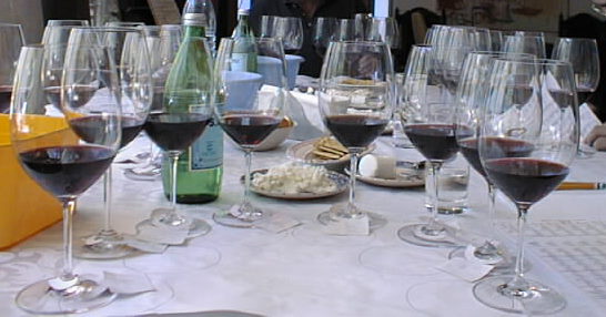
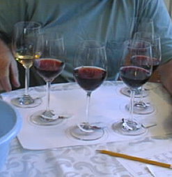
Hold the wine glass by the stem or base and not the neck or the bowl. Start by holding the glass toward a light source or a white background and tilt it around at an angle.
When looking at wine, we look for three main characteristics: color, depth and clarity
In wine we will see different shades and hues of Reds Yellows or Pinks, (in red white or rose’ wines), these may appear either diluted or deep, they may appear radiant or dull, even cloudy or hazy, all of these are indicative of the wine age and quality and will be discussed later in details.
For now, in general, when we look at red wines for instance, a brilliant red color usually indicates a wine in its prime, a purplish hue may indicate a very young wine and a brown hue may indicate that a wine is slightly oxidized or been lying down for quite a while, or even past its prime.
 Isaac Newton, in his theory of color, observed that color is not inherent inside objects, but rather, the surface of an object reflects some colors and absorbs all the others. We perceive only the reflected colors. So, red is not “inside” the liquid of red wine. It is the surface of the wine that reflects the wavelengths we see as red and absorbing all the rest. (An object appears white when it reflects all wavelengths and black when it absorbs them all.)
Cranial Nerves associated with wine tasting
Our first encounter with wine is through the sense of sight.
Cranial Nerve II – The Optic nerve is a pure sensory nerve which supplies the photoreceptor cell of the retina at the back of the eye ball, basically it allows us to see shapes, colors, hues, clarity and depth, all perceived through the eyes (as far as the wine in the glass goes). You will also be able to see the bottle, shape and color, the label with all the information regarding the wine, region, even sub region, pedigree, wine maker, vintage year etc. You can “scan” your company and the surrounding of your wine experience. Sight gives us only certain clues regarding the wine before us they are mainly initial clues regarding the wine’s condition, age, freshness (according to the grape variety), some of these “clues” will have to be reassessed in combination with the other senses smell, taste and touch.
In order to see sideways, up or down, you need to “use” another 3 of the cranial nerves which are pure Motor nerves they initiate voluntary movement of the eye and lids. Cranial nerves III, IV and VI, which together, control the six muscles of the eye, the eyeball and eye lid movement.
Together these 4 (out of 12 Cranial nerves) facilitate vision.
Seeing is believing, the neurophysiology of Vision
 Cranial nerve II: Optic nerve
The optic nerve is composed of axons of the ganglion cells in the eye. It carries visual information to the brain. This is a pure sensory nerve fiber. This nerve travels from the back of the eye ball, entering the brain through the orbit at a small “hole” (the optic canal) in the skull bone. The 2 Optic nerves one for each eye, meet & cross each other to form the optic chiasm. (Right eye vision is partially perceived on the left side of the brain and vice versa. The brain does not receive signals from each eye unilaterally. Half of each optical field is directed to the opposite part of the brain. This occurs when the bundled fibers of the optic nerves meet and cross at the optic chiasm (cross road), located just a few centimeters inside the brain. It runs to the vision center of the brain – the Visual Cortex, here, information is interpreted and true vision is formed.
The eye is the sense organ with all its part Cornea Lens Iris Retina and behind them specialty receptors sending chemical and electrical signals to the brain for interpretation through a pipe called the Optic nerve.

Cranial nerve III: Oculomotor nerve
The Oculomotor nerve is composed of motor axons. This is a pure motor nerve. It provides somatic motor innervations to four of the eye muscles which allow movement of the eyeball. It also innervates the muscles of the upper eyelid and the inner eye muscles that control the amount of light that enters through the pupil. (The pupillary eye muscles)
Cranial nerve IV: Trochlear nerve
The Trochlear nerve provides somatic motor innervations to one of the upper eye muscles it controls the downwards and sideways movement of eyeball, helps you see where your wine glass before you pick it up or alas spill the above wines (and many others) on the white table cloth!!. It is also a pure motor nerve fiber.
Cranial nerve VI: Abducens nerve
The Abducens nerve carries somatic motor innervations to one of the outer eye muscles, it controls the eyes side movement, careful who’s sitting next to you, who sneaks a hand towards you glass during conversation with the person next to you!!! It is another pure motor nerve fiber.
The anatomy of the eye


The Cornea: The cornea is a round, transparent dome that acts as the outer window of the eye. It is the structure that focuses the light that enters the eye. It comprises five parts. All the parts work together to protect the eye and help in the proper working of the cornea as a whole.
The Lens: The lens is that part of the human eye that is located immediately behind the iris. It is transparent, elastic and crystalline. Its role is to focus the light and move towards the retina.
The Iris: The colored part of the eye is known as iris. It is present in the eye in the form of a thin diaphragm. The iris lies between the cornea and the crystalline lens. The color is due to the presence of a pigment. It is the iris that gives your eyes a particular color. The basic iris colors are blue, green and brown. Majority of humans have varying shades of these colors. It is composed of connective tissues and smooth muscle fibers. The composition of the iris enables it to dilate or contract the pupil, which in turn controls the amount of light that falls on the retina.
The Pupil: The hole in the center of the eye through which the light passes, is called the pupil. The pupil gets bigger and smaller depending on the amount of light falling on the eye.
The Sclera: The sclera is the whitish, opaque part of the eye, which is connected to the cornea. Its role is to provide protection and meet the purpose of attachment for the muscles that enables eye to move.
The Vitreous Humor: It is the jelly like substance that is present within the interior chamber behind the lens. It is that part of the human eye whose role is to provide pressure inside the eye and keep it inflated
The Retina: The retina is the innermost layer of the eye. It consists of nerve tissue that senses the light entering the eye. Its function is to send impulses through the optic nerve back to the brain, where it gets translated into the images that we see. There are four types of light-sensitive receptors present in the retina.. The retina is considered to be part of the brain itself, it is covered by millions of light-sensitive cells, some shaped like rods and some like cones. These receptors process the light into nerve impulses and pass them along to the cortex of the brain via the optic nerve.
The Fovea: The Fovea is a part of the eye, located in the center of the macula region of the retina. The fovea is responsible for sharp vision which is necessary when visual details are most important.
The Optic Nerve: The continuation of the axons of the ganglion cells in the retina is known as the optic nerve. It connects the eye with the brain. The optic nerve emerges from the back of the eye, travels through the skull and stops inside the skull bone, and ends up at the back of the brain. This part of the brain is known as visual cortex. It is responsible for receiving information from the eyes and interpreting it.
Now we can see, next post I’ll try to figure out how we can actually see, what we see and why????????????
YOUR WINEGUIDE
some images from http://www.positscience.com/brain-resources/image-gallery/vision-images
